How To Diagnose Heart Disease?
The diagnosis of heart disease is a task reserved for health professionals, through additional examinations designed for this purpose. What are the methods used? Discover them now!

A cardiac pathology concerns the area of the heart. Unlike cardiovascular disease, which also includes arteries and veins. In addition to arrhythmia, there are other examples such as dilation of the heart muscle, infarctions and heart failure. When the pathology affects the internal valves, we can cite stenosis and prolapse.
We will use the same complementary methods for the majority of these disorders. The diagnosis of heart disease usually involves following the steps of a protocol that involves several examinations, from the simplest to the most complex. We detail them here.
What tests can diagnose heart disease?
As mentioned above, the complementary methods for diagnosing heart disease are the same for several pathologies. An electrocardiogram makes it possible, for example, to detect an arrhythmia and an infarction, although these pathologies are different.
Electrocardiogram
An electrocardiogram (ECG) is an electrical recording of the activity of the heart muscle. Thanks to electrodes placed outside the body, we can perceive the electrical variations emitted by the beating of the heart. They originate from the internal system, which contains heart tissue and controls the beat.
The test does not require anesthesia or special preparation. It is even carried out on an outpatient basis and the patient can leave after half an hour. Sometimes it is a routine check to follow any changes in the situation. In other cases, these are checks requested after an already established pathology, such as a heart attack or arterial hypertension.
The recording is materialized by the measuring device in the form of a document with a line marking the beats. The interpretation of the test depends on the training of the doctor. There are already established reading protocols that indicate the meaning of each line drawn.

Echocardiogram
To diagnose heart disease, an echocardiogram may be used. The test consists of an ultrasound, like the one done to check a pregnancy. In this case, it is intended for the heart.
Echo-like signals are sent through a device called a transducer, which bounce off the heart muscle and return to be interpreted as an image by a monitor. One can then directly see the movement, the beats, the form and the cardiovascular dynamics.
A variation is Doppler , which adds colors to the image to distinguish venous blood from arterial blood. Nowadays, this modality is almost always used because this information is crucial.
Stress test
The technical name for the stress test to diagnose heart disease is “ergometry”. In simple terms, it is about stimulating the heart through physical exercise and recording what happens under stress. EKGs and echocardiograms are therefore performed while the patient is running on a treadmill or pedaling on an indoor bicycle.
Certain parameters are taken into account during a stress test so as not to exceed the limits that could endanger the person performing the test. We set limits for the heart rate, for the incline of the treadmill if it is mobile, and we pay attention to signs that could stop the effort.
It is a very useful complementary method for simulating everyday situations. Times when patients are forced to run or jog, or climb the stairs. We thus study the heart by reproducing reality, but in a controlled environment.
Cardiac Holter
This test, which doctors call holter for short, is actually a long-lasting EKG. Through a device that patients use for one or two days, the electrical activity of the heart is recorded .
Later, through computer programs, variations such as the number of arrhythmias, accelerations, tachycardia and bradycardias are counted. The advantage of this device is that it makes it possible to monitor situations that escape the ECG, such as the patient’s sleep phases. This then makes it possible to diagnose heart diseases that hitherto escaped testing.
Cardiac catheterization
A cardiac catheterization is a surgical procedure that involves the insertion of a catheter into the circulatory system. Access is often through the upper or lower limbs, until reaching the heart with the device, which can take measurements or inject X-ray dyes.
Usually, the procedure relies on external images that will follow the appearance of the x-ray dye. In this way, one can see the heart chambers in sufficient detail to detect dysfunctions and anatomical failures.
Not all patients can undergo catheterization, but those for whom it is indicated may benefit from treatment at the same time. Once the catheter is inserted, it is possible to make repairs or remove clots that block the coronary arteries, for example.
Cardiac magnetic resonance
Advances in medical imaging have led to the development of a specific magnetic resonance (MR) for the heart. From the same functioning of MR for the rest of the body, this time a non-irradiating force is used to observe the heart muscle. The indications obtained may be superimposed on those of the echocardiogram.
Computer Assisted Cardiac Tomography
Computed Axial Tomography (CAT) takes the fundamentals of TACO of the body, but is only applied to heart tissue. It uses x-rays, like x-rays, to generate images of the chest and provide important information. The patient lies down on a table which is inserted into a tube through which the x-rays will be emitted.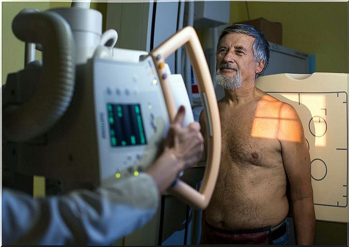
Reduce the risk of heart disease
Beyond diagnosing heart disease, it’s important to prevent it. Current methods do not allow early detection of cardiac damage that is dangerous to health. But proper diet, exercise, reducing stress, and regular check-ups are the best tools to avoid them.
- In terms of diet, it should be remembered that it has been proven that favoring vegetables over meat, and including natural, unprocessed foods rich in omega-3, helps protect cardiovascular health.
- With regard to physical exercise, scientific studies agree that a regular sports session, lasting between 30 and 60 minutes, is sufficient to reduce the risk.
- To reduce stress, there are various techniques ranging from deep breathing to yoga, meditation and mindfulness. Each person has an affinity for one or the other depending on their personality and cultural background.
- Finally, regular checks with medical professionals are essential, at least once a year. Adults are recommended to have one EKG per year, although people with diabetes and high performance athletes should do them more frequently, every 6 months.
If all this is not enough, it will be necessary to perform an additional method among those listed to arrive at the correct diagnosis. If in doubt, it is best to see a doctor, especially if there are warning symptoms.